
Zudena
D. Warren Spence, MA
- Sleep and Alertness Clinic, University Health
- Network, Toronto, Ontario, Canada
The etiology new erectile dysfunction drugs 2014 buy cheapest zudena, evaluation erectile dysfunction therapy treatment cheap 100mg zudena with mastercard, particularly during growth erectile dysfunction doctor london buy discount zudena 100mg line, and the management of these challenging entities will Families of children with craniofacial anomalies present many be presented erectile dysfunction what kind of doctor order 100mg zudena with amex. Crerand latest erectile dysfunction medications order zudena 100 mg free shipping, PhD issues will be discussed and cases will be presented to amplify the discussion impotence natural treatment clary sage cheap zudena 100 mg fast delivery. Conference attendees will develop an the experience of having a facial disfigurement can present understanding of the challenges faced by the primary care numerous challenges for children and their families. Facial provider, the importance of communication among everyone disfigurement can affect not only how children view involved in the patients care and each persons role in themselves but how they are perceived of and treated by providing a medical home for the child with a craniofacial others. This presentation will examine factors that impact psychosocial adjustment to facial disfigurement with Timely and appropriate dental evaluation and management is an emphasis on body image and its role in psychosocial a key foundation for successful craniofacial team management functioning and quality of life. Intervention strategies that can of children with asymmetries and other craniofacial anomalies. Rare craniofacial conditions, including Craniofrontonasal or Attendees should also have an understanding of the range of Fibrous Dysplasia, Vascular Anomalies, Beckwith-Wiedemann strategies which integrate dental management approaches Syndrome and Hemifacial Atrophy, present significant into the overall team care plan. The nurse/team coordinator is often their air processes involved in the orthodontic and dental care of traffic controller the lifeline between the patient/family and individuals affected by facial asymmetry problems. Paramount all who participate in their care, giving us a unique to providing ideal care to these patients is the presence of a perspective. This presentation will highlight the road that we good dentition, and the role of all Team members in this all travel together, guided by our primary goal of helping our regard will be reviewed. Also dependent on a good outcome is patients to become an accepted and productive member of a proper diagnosis and treatment plan, not a simple society. Lastly, examples of the clinical means by which dental professionals can contribute to the correction of these asymmetry problems will be provided. She is also Assistant Professor of Clinical Plastic Surgery, Speech and Hearing Science, and Pediatrics at the Ohio State University. Her clinical and research interests include perceptual and instrumental assessment of velopharyngeal dysfunction in children with cleft palate, craniofacial anomalies and 22q11. Bradleys research focuses on basic science studies and translational research related to bone biology, bone tissue engineering, and wound healing. In clinical outcome investigations, the research team focuses on refining surgical protocols and innovative surgical procedures. All of these studies aim to improve surgical outcomes for patients with craniofacial syndromes. She is a neurodevelopmental pediatrician and geneticist and serves as consultant to the Craniofacial Program. Bull is a frequent speaker nationally and internationally and has served on the American Academy of Pediatrics Committee on Genetics and is currently on the Board of Directors. Pat received the Donna Pruzansky Memorial Fund Award from the Cleft Palate Foundation in 1992, and since that time, has been a very active nursing member of the American Cleft Palate-Craniofacial Association. She previously served as a craniofacial team psychologist at the Childrens Hospital of Philadelphia and was an Assistant Professor in the Department of Surgery at the University of Pennsylvanias Perelman School of Medicine. Drake, a pediatric otolaryngologist, has clinical and research interests which focus on pediatric airway disorders and craniofacial anomalies. She has been named on both Americas Top Doctors and Americas Best Doctors lists for many years. Tucker Award, for significant contributions to the field of pediatric laryngology, from the American Laryngological Association. He completed his plastic surgery residency in the Harvard Training Program, followed by a craniofacial fellowship at Boston Childrens Hospital. He is a member of the Vascular Anomalies Center, Co-Directs the Lymphedema Program, and Directs the Department of Plastic Surgery research laboratory. His clinical and research focus is in the fields of vascular anomalies and lymphedema. He then received his medical degree and general surgery training at the University of Connecticut. Fellowship training in CranioMaxillofacial Surgery was abroad at the Royal Childrens Hospital in Melbourne, Australia and Alder Hey Childrens Hospital in Liverpool, England. He has had academic appointments at the Universities of Connecticut, Maryland, Minnesota and currently is Associate Clinical Professor of Surgery at the West Virginia School of Medicine. He is very active in the American Cleft Palate-Craniofacial Association as well as his specialty organizations. She has served the Association in many capacities, most recently as Chair of the Committee on Accreditation of Teams. She is interested in normal and abnormal morphogenesis as well as the the etiology and pathogenesis of cleft and craniofacial disorders. She has written numerous professional articles and 22 book chapters in speech pathology and medical texts. She is the author of the book entitled Cleft Palate and Craniofacial Anomalies: the Effects on Speech and Resonance, 2nd edition (Delmar Cengage Learning, 2008). She is an oral & maxillofacial surgeon with expertise in craniofacial anomalies, orthognathic reconstruction, and benign bone tumors, such as fibrous dysplasia. Marsh received the Hopkins Alumni Associations Knowledge for the World award in 2011 for his international cleft care volunteerism. Mooney, PhD, is Professor and Chair, Department of Oral Biology, University of Pittsburgh with appointments in Anthropology, Plastic Surgery, Orthodontics, and Communication Sciences and Disorders. Mooneys research interests include factors that affect growth and development and the development of animal models of craniofacial. After teaching 10 years at the Universities of Indiana and North Carolina, he became the first Director of Orthodontics at Arkansas Childrens Hospital, providing all orthodontic services for their cleft, craniofacial and special needs patients. He has contributed multiple chapters on orthodontic treatment of patients with cleft/craniofacial conditions. He is an Affiliate Professor in the Departments of Pediatric Dentistry and Orthodontics at the University of Washington and a member of the active medical staff at Seattle Childrens Hospital and Swedish Medical Center. After 16 years as an active member he now continues to have a strong working relationship with the Craniofacial Center at Seattle Childrens Hospital. Forty years worth of reconstructive procedures followed, but Verdis craniofacial condition didnt prevent her from being an activist on behalf of others. Sharp, PhD, Western Michigan University, the 2014 Program Committee is pleased to announce the Dept. What makes some teams excel while others fail separate freestanding educational programs. What separates great teams from groups of evolved over the years and is now focused on patient, and individuals that struggle to reach their full potential This Fundamental Laws of Team Care will discuss the basic presentation will highlight the major services, projects and principles and strategies that are essential to building a products of the Cleft Palate Foundation and its mission. Understanding and applying Particular emphasis will be placed on how it supports team these ideas, including the Law of Significance (Yes, It Takes a care, and a description of the foundations educational Team), the Law of Purpose (It Really is the Vision Thing), and materials and programs including student scholarships for both the Law of the Helm (The Team Sinks or Sails on Leadership), undergraduate and graduate specialty education. In addition, will not only help your team fulfill its mission but also the types of research funding available will be described. Learning and Attendees following this presentation will be able to discuss practicing these laws will enhance your capacity to unlock your the basic mission and function of the Cleft Palate Foundation. This presentation will provide a review of clinical study designs, highlight International Congress last May, reports considerations for participation in research, and offer tips to from forum leaders indicated a wide get started. This is your Palate Clinic opportunity to meet and greet Internal audit of our clinical outcomes is an obligation we have colleagues in your discipline from as part of quality assurance and improvement. Forum specialties measure outcomes of significance, an additional opportunity presents itself for intercenter outcomes comparisons and and room assignments are: research. Such collaboration can provide insight into the processes and outcomes of treatment or comparable services elsewhere and the exchange of clearly successful practices. We will explain the importance of meaningful Michael Friel, Sunil Tholpady, Robert J. These benefits will topics and information was exchanged, but even more translate into improved care for the children and communities importantly, connections were made. Panel speakers will explain the pathways to opportunity to meet and greet colleagues in your leadership development within the organization and will elicit discipline from around the world! See room assignments audience participation as we undertake a voyage in leadership on Summary of Events, Page 128. Finally, patients with common Craniofacial B=Beginner L=Lecture syndromes that may have coexisting speech deficits. I=Intermediate H=Hands-on Learners will be able to identify the key components of A=Advanced P=Panel a speech therapy home program. Participants will have an related to cost and time, selecting valid and reliable understanding of genetic contribution to nonsyndromic tools, and equipment. Inheritance patterns and Anna Thurmes, Kelly Nett Cordero, Kristina Wilson, current technologies for genetic testing will be Adriane Baylis, Kathy Chapman, Angela Dixon, Cindy highlighted. Specific cases will be presented to Dobbelsteyn, Debbie Sell, Judith Trost-Cardamone emphasize the value of incorporating genetic Room: Utah counselors and geneticists to improve the overall healthcare provided by the interdisciplinary team. Short video clips of these techniques will Strategies for design and planning of a nurse led be presented for clarity. There will be a discussion of multicenter research study, an overview of the methods for achieving carry-over once normal background, purpose, research questions, methods, production is achieved. Participants will receive a plan for analysis of a proposed multicenter study and handout of techniques, including those that can be proposed data collection tool will be discussed. Typical Room: Austin/Boston scenarios will be presented and recommendations will be made regarding the purpose, content and structure of a prenatal counseling session. Mark Mooney, Joseph Losee Objective: Attendees will be able to identify and discuss three research questions related to cleft and craniofacial care. Objective: Participants will be able to Cleft will be presented, with assessments from clinicians, identify at least two common challenges for new mental patients and caregivers. Deidrick, Sandra Sinclair, Heather Snyder Hillary Broder, Margot Stein, Canice Crerand, Cynthia Room: Marriott 7 Cassell, John Riski C. This course will provide the audience with a detailed description of the use of Nasoalveolar Molding Technique Codes: Instruction Level Format for infants born with unilateral and bilateral cleft B=Beginner L=Lecture deformities. The course will also discussed maxillary arch I=Intermediate H=Hands-on preparation for a secondary bone grafting procedure during A=Advanced P=Panel the mixed dentition. Educational Objective: the participant V=Varied R=Roundtable should be able to introduce the discussed principles into his/her daily orthopedic/orthodontic practice. Demonstration and hands-on the practicing orthodontist and surgeon who treats experence with specific cleft feeders will be provided. We Finally, feeding issues specific to cleft related will focus both on orthodontic and surgical challenges. Dailey, Brandon Viet, Kerry Mandulak with residual fistulae, segmental osteotomies, Room: Santa Fe simultaneous bone grafting, management of existing posterior pharyngeal flap, and impact on sleep apnea B. Final considerations of well as determining which services are feasible to orthodontic finishing will be discussed as well implement, given clinical demands and available Anand Kumar, Lindsay Schuster, Derek Steinbacher resources. For each condition, participants will: 1) this course will use an interactive format to review define diagnostic criteria, differential diagnoses, and common procedures offered to patients with cleft lip confirmatory studies, 2) identify health concerns that and palate. Procedures reviewed will include cleft lip could impact readiness for surgery or increase risk for repair, cleft palate repair, pharyngeal flap and sphincter adverse outcome, and 3) provide critical appraisal of a pharyngoplasty. Three dimensional Emily Gallagher, Ophir Klein, Robert Byrd, Katrina understanding will be emphasized. Correlations will be Dipple, Charlotte Lewis, Michael Cunningham drawn between specific techniques and the theoretical Room: Austin/Boston advantages and disadvantages among them. This masters class will provide a comprehensive overview of the multifactorial K.
The most current recommendation is published in the Red Book 2003 of the American Academy of Pediatrics erectile dysfunction treatment garlic purchase 100mg zudena otc. These infants will also benefit from receiving influenza immunization at 6 months chronological age during the cooler winter months (3) erectile dysfunction doctors huntsville al order zudena 100mg line. The premature infant is ready for discharge when he/she is able to fulfill the following criteria: 1) ability to appropriately regulate their temperature without the need for technological support erectile dysfunction doctors jacksonville fl purchase zudena amex, 2) ability to ingest adequate calories to achieve consistent growth erectile dysfunction commercials buy genuine zudena on-line, and 3) to have demonstrated other parameters of global physiologic stability (the absence of clinically significant apnea erectile dysfunction at 55 discount 100 mg zudena fast delivery, bradycardia zocor impotence cheap zudena 100 mg overnight delivery, or hypoxemia). Thus, the process of discharge of the infant is a continuum that begins several days to weeks prior to the actual discharge of the infant. In addition, these infants are frequently born into families who are already high-risk. On a positive note, if an optimal nurturing environment is provided, there is evidence to suggest that it can result in a significant improvement in overall long term outcome. True/False: Morbidity associated with prematurity is a significant contributor to the infant mortality rate. Keeping the delivery room warm and performing the stabilization under a preheated radiant warmer. They are born with inadequate glycogen stores and have immature homeostatic mechanisms to mobilize glucose. Feeding difficulties in premature infants are usually secondary to (choose one): a. Obstructive secondary to collapse of the upper airway structures and closure of the glottis. True/False: the weight of the premature infant is an absolute criterion for discharge from the hospital. His face is symmetrical with normal palpebral fissures, normal red reflexes, patent nares, normal ears, no clefts, and no neck masses. His abdomen is soft and round with normal bowel sounds, no masses and no organomegaly. The constellation of signs and symptoms can be the result of pulmonary, cardiac, metabolic, infectious, renal, gastroenterological and neurologic pathologic processes. Newborns with disorders involving any one of these organ systems may present with varying degrees of tachypnea, retractions, grunting, cyanosis, lethargy and tachycardia. Given the similar presentations, the circumstances of the newborns birth provide important clues to the diagnosis. The chest radiographs reveal hyperinflation with clear lung parenchyma except Page 91 for perihilar linear densities and fluid in the fissures. The pathophysiological mechanism is the delayed resorption of fetal lung fluid which eventually clears over the next several hours to days. In addition, while many infants have the onset of symptoms at birth, some infants have an asymptomatic period of several hours before respiratory distress becomes apparent. The pathophysiologic mechanism is the obstruction of large and small airways with the aspirated material (meconium, blood, amniotic fluid contents). The duration of distress with mild to moderate aspiration syndromes is from several hours to days. A large randomized trial has confirmed that aggressive intubation is not necessary for most infants with meconium in the amniotic fluid. In addition to respiratory distress, a severe air leak condition may cause hypotension (due to decreases in cardiac output), muffled heart tones, abdominal distention, asymmetric chest shape and deviation of the cardiac sounds. Chest radiographs are diagnostic with free air in the hemithorax and a visible edge of the collapsed lung. Treatment of significant air leak syndromes requires immediate air evacuation (thoracentesis or pericardiocentesis) with a needle or small catheter, followed by chest or pericardial tube insertion. Most infants are less than 34 weeks gestation and the incidence and severity increase with decreasing gestation age. These premature infants have progressively more severe respiratory distress after birth. The presence of apnea suggests severe disease accompanied by refractory hypoxemia and acidosis. Without surfactant, the surface tension of the alveolar sacs is high, leading to an increased tendency of the alveoli to collapse. This leads to the network of air-filled alveoli juxtaposed to atelectatic alveoli and creates the reticulogranular pattern (ground glass appearance) of the lung. Grunting is the infants attempt to maintain the pressures and gas volume within the lung by causing expiratory braking using the vocal cords (the glottis is partially closed during exhalation to maintain alveolar distending pressure during exhalation). Today, several types of animal based surfactants have been approved for clinical use. Page 92 Moderately premature infants (29 to 34 weeks gestation) are usually extubated within several days after treatment. However, extremely premature infants (23 to 28 weeks gestation) may continue to require positive pressure respiratory support for several weeks. Mothers with intrapartum fever and prolonged rupture of membranes (>18-24 hours) have a greater risk of transmitting infections to their infants. Due to the serious consequences associated with delays in treatment for infections, many infants with non-infectious conditions are evaluated and empirically treated with antibiotics for this possibility. A left shift with greater than 20% band forms of the total neutrophils is suggestive of infection as are neutrophilic vacuoles and toxic granulation. Supportive care may include mechanical ventilation, supplemental oxygen, inotropic agents for hypotension and nitric oxide for infection associated pulmonary hypertension. Most infants with cyanotic heart disease typically have a paucity of respiratory distress symptoms except for cyanosis or duskiness. Typically the chest radiograph reveals a normal sized heart or cardiomegaly with clear lung fields and decreased vascular markings (due to diminished pulmonary blood flow). When the infant develops respiratory symptoms, it is usually from severe hypoxemia or acidosis. The infant who is cyanotic with respiratory distress and does not respond to supplemental oxygen. Infants with both cyanosis and respiratory distress may have chest radiographs typical of pulmonary disease. Therapy for cyanotic heart disease consists of medical support until definitive surgical repair can take place. Multistaged open heart surgery may be necessary for most complex cyanotic heart diseases. Bowel sounds are heard over the chest if air enters the intestines from spontaneous breathing or mask valve ventilation. The chest radiograph reveals a bowel gas pattern typically in the left hemithorax with a mediastinal shift to the right. High frequency ventilation and nitric oxide therapy are used to treat the bilateral hypoplastic lungs. The hypoplastic lungs develop excessive and abnormal musculature of the pulmonary vessels which lead to pulmonary hypertension. However, based on the time of onset and the progression and severity of the symptoms, other causes of respiratory distress must be entertained. In the case presentation at the beginning of this chapter, the later onset of respiratory distress which increases in severity with time, suggests either aspiration or an infectious process. Cyanotic congenital heart disease: Clinical factors: Heart murmur, persistent hypoxia despite supplemental oxygen. These conditions lead to a reticulogranular infiltrate (ground glass) and air bronchogram pattern on the chest radiograph. At 5 hours of age, with the second feeding, the baby appears tachypneic and cyanotic, and he is therefore taken to the nursery for further evaluation. His heart is regular with a grade 2/6 systolic ejection murmur at the lower left sternal border. The infant is mechanically ventilated and subsequently transported to a pediatric cardiac surgical specialty center. The newborn infant with cyanosis challenges the clinician to identify the cause and institute appropriate treatment. As with all neonatal conditions, diagnosis is aided by obtaining a thorough maternal and birth history. Clues to infant problems may be found in pregnancy screening tests such as maternal serum alpha-fetoprotein, a marker for fetal aneuploidy, or knowledge of preexisting maternal medical conditions such as diabetes. Maternal serologies and cultures identify newborns at risk for perinatal group B streptococcal pneumonia or intrauterine toxoplasmosis infection. The progress of labor and delivery, as reflected in Apgar scoring and delivery room resuscitation, also provides valuable information. Fetal heart rate pattern abnormalities, meconium staining of the amniotic fluid, maternal fever or bleeding may suggest neonatal pneumonia, hypoxic-ischemic injury, meconium aspiration syndrome or persistent pulmonary hypertension. Peripheral cyanosis (acrocyanosis) is a normal finding in newborns and does not indicate systemic desaturation. Pigmentation of the vermilion border and facial bruising may also masquerade as cyanosis. Likewise, the polycythemic infant with a normal oxygen saturation may appear cyanotic from peripheral sludging of desaturated red cells despite normal oxygen saturation. Only later is the hypoxia detected with the investigation of ancillary signs such as tachypnea, tachycardia or other signs of distress. The quality and symmetry of breath sounds may suggest focal disorders such as pneumothorax and diaphragmatic hernia or more generalized ones such as respiratory distress syndrome. More infrequently heard holosystolic or diastolic murmurs require definitive evaluation. Conversely, many serious cyanotic congenital heart malformations are not accompanied by murmurs. Generally weak pulses denote systemic hypoperfusion as in low volume states and decreased cardiac output. The association of cyanosis with dysmorphic features may provide diagnostic information. Facial and limb deformation associated with oligohydramnios is associated with hypoplastic lungs and pulmonary hypertension leading to cyanosis (5). Page 95 the most common congenital heart lesions presenting with cyanosis in the newborn period are those of the hypoplastic right heart syndrome complex (pulmonary and tricuspid atresia) and transposition of the great vessels. The basic pathophysiologic mechanisms leading to hypoxemia are inadequate perfusion of the lungs or marked right-to-left shunting and admixture of desaturated venous blood in the systemic arterial circulation. Less common conditions include congenital anomalies of the lungs such as congenital diaphragmatic hernia, tracheoesophageal fistula and pulmonary hypoplasia. Transient tachypnea of the newborn, a common neonatal respiratory disorder, generally is not accompanied by marked cyanosis. Central nervous system dysfunction caused by hypoxic ischemic injury, seizures, intracranial hemorrhage, infection, or metabolic derangement such as hypoglycemia may lead to cyanosis. An usual pattern of methemoglobinemia has also been described in infants with diarrheal disease of various etiologies including milk protein intolerance and infectious gastroenteritis accompanied by severe systemic acidosis (3,4). However, prior to obtaining a cardiology consultation and echocardiogram, the clinician may perform a number of other valuable tests to define the cause or mechanism of cyanosis. An anteroposterior chest x-ray will identify pneumonia, pneumothorax or the intrathoracic bowel gas patterns characteristic of diaphragmatic hernia. The classic cardiac silhouettes of transposition of the great vessels ("egg on side"), total anomalous pulmonary venous return ("snowman" heart) and tetralogy of Fallot (boot-shape) are uncommon in the newborn period. Two pulse oximeter probes placed simultaneously on an upper and lower extremity will give clues to right-to-left shunting across a patent ductus arteriosus. Likewise, a marked differential in paO2 between blood drawn from an upper extremity artery and umbilical artery catheter or posterior tibial artery carries the same implication. Rarely is cardiac catheterization required, except in confusing cases of complex anatomy or instances of uncertainty. Targeted treatment is dependent on accurate diagnosis and understanding of pathophysiology. Oxygen, a potent pulmonary vasodilator, may increase pulmonary blood flow at the expense of systemic perfusion. In anomalous pulmonary venous return with obstruction, oxygen therapy may be particularly hazardous contributing to increasing pulmonary venous hypertension and clinical deterioration. Anticipation of this common complication and stabilization of the patients airway and ready availability of ventilatory support can avoid deterioration especially in the transport setting. In transposition of the great vessels, timing and severity of presentation relates to the degree of right/left mixing. If there is a coexisting ventricular septal defect with adequate mixing, recognition may be delayed up to several weeks. Red blood cell transfusion is commonly employed to support oxygen carrying and delivery capacity. Attention to fluid and electrolyte balance includes calcium maintenance for optimal cardiac performance. Acid-base derangement is addressed with attention to treatment of underlying disorders and the judicious use of sodium bicarbonate. Examples are the arterial switch (Jatene) procedure for transposition of the great vessels and primary repair of anomalous pulmonary venous drainage and tetralogy of Fallot. The best outcomes for neonatal cardiac surgery are seen in pediatric cardiac centers with high volumes and skilled teams (8,11). What are the 2 most common congenital heart diseases leading to cyanosis in the newborn period A 2 day old term infant previously thought to be well and about to be discharged from the nursery becomes acutely pale, slightly cyanotic, with weak femoral and brachial pulses. Hawaii Birth Defects Program 1986-1998 Statewide Data, Surveillance Report Number 7 on Birth Defects in Hawaii, January 1,1986-December 31, December 1999, 1-126. Dietary protein intolerance in infants with transient methemoglobinemia and diarrhea.
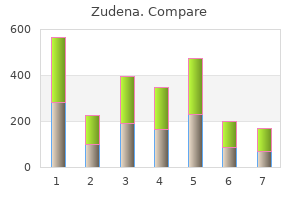
Proven zudena 100 mg. Can Erectile Dysfunction Be Cured - Curing ED (Impotence) is Not That Hard.
State law and provider contracts with insurers provide for appeals and one should be filed if it is felt that a patients claim or your bill was unfairly denied erectile dysfunction aids purchase zudena toronto. In short erectile dysfunction massage techniques order zudena 100 mg otc, documentation of the services rendered and a thorough knowledge of coding rules and procedures are essential to receiving the best compensation for your services erectile dysfunction by age cheap zudena line. Provider contracts with third party payers include provisions for fee schedules to be established new erectile dysfunction drugs 2014 buy zudena toronto. You may charge whatever you wish for a given service antihypertensive that causes erectile dysfunction order zudena, but the insurer will pay no more than the "maximum allowable charge" (the amount for a service listed on the fee schedule) for the service male erectile dysfunction icd 9 discount 100mg zudena fast delivery. For example, it may pay 80% of an office visit (the actual percentage varies with different plans). This intermediary may choose capitation or a fee for service method of paying the physicians who render the care. Both providers and insurers are expected to provide those medically necessary services authorized by the terms of the physicians contract and the patients coverage. It is important to know that an insurance policy rarely covers all possibilities, but rather only those services that are "covered services" and are "medically necessary" (a term that is defined in state law) will be paid for. The sample case at the beginning of the chapter is another example, such that developmental speech abnormalities were not a covered service in the patients plan, even though the need for the service is appropriate. Some community obligations exist, such as not abusing antibiotics and causing the development of resistant organisms, but the basic obligation is to the individual patient. Third party payers must consider their total membership and the community as well as the individual member. Prices must be affordable for those paying the premium and insurers have an obligation to remain fiscally solvent through the terms of their contracts. The provider, whose attention is called to denials and not to the majority of claims that are immediately paid, may feel that all the insurer does is try to cheat them. Conflict resolution must occur with each side appreciating the others role in the overall scheme of managing patient care. True/False: A charge is adjusted downward because it exceeds the maximum allowed for that service. True/False: Due to their large reserves, insurers have minimal budgetary constraints in spending. The patient must be informed beforehand that the service may not be covered and that he or she will be expected to pay if they wish to have the service done. Contracts between third party payers and providers stipulate that balance billing is not allowed when fees exceed maximum allowable charge on a covered service. An insurer must observe its operating budget, which is dependent on the premiums received. Repeated withdrawals from reserves threaten the solvency of the third party payer. In Hawaii, the latter includes childhood preventive health services and immunizations through age five years. Insurers can abuse downcoding if it is done arbitrarily or solely to pay the provider less. A system of disease classification based on work by the World Health Organization and issued in the United States by the U. Managed Care: A means of providing health care services within a defined network of health care providers that is given the responsibility of managing utilization of health care services and providing quality, cost-effective health care (1). Medicaid: A jointly funded, Federal-State health insurance program for certain low-income and needy people. Third party payers changed this to refer to care that their payment teams deemed necessary for the management of a given patient. Other contractual requirements may include mandatory participation in aspects of the plan to monitor quality or to save money, such as following a formulary. Upcoding is appropriate if a higher level of service was actually rendered; inappropriate if it was not. She typically feeds him at night, and he refuses to go to sleep without a bottle of milk or apple juice. Intraorally, there are opaque brown and grey specks on the enamel surface of several upper primary teeth. Moderate lesions are visible in the upper canines, upper first molars, and lower first molars. His mother is told to wean him off the bottle and to brush his teeth at least twice a day. Pediatricians and primary care family physicians play a vital role in promoting good oral hygiene as a life long habit that begins during infancy. They are the first and most frequent health care providers seen by infants and young children, during the formative years of oral health care. The primary structures of the tooth are the: 1) enamel (outermost protective layer), 2) dentin (calcified tissue layer deep to the enamel), 3) gingival margin (region of the gum surrounding the tooth), 4) pulp (soft tissue at the core of the tooth which contains blood vessels, nerves and lymphatics), 5) cementum (layer of bony tissue covering the tooth root surface), 6) periodontal ligament (membrane around tooth attaching it to alveolar bone), 7) alveolar bone (bone that surrounds the root and forms the socket for the tooth), 8) neurovascular bundle (nerves, arteries, and veins in the dental pulp that exits at the root of the tooth). There are 20 primary teeth (described by positions A through E) and 32 permanent teeth (described by positions 1 through 8). From the center proceeding posteriorly: central incisor (#A, #1), lateral incisor (#B, #2), canine (#C, #3), first premolar (#4), second premolar (#5), first molar (#D, #6), second molar (#E, #7), third molar (#8). The formation of human dentition begins as early as the 6th week in utero, during which, tooth buds of the primary (deciduous) teeth develop at 10 specific sites in the developing maxilla and mandible (1,2). Primary teeth begin to calcify at about 3 to 4 months in utero, and the enamel of all crowns is completed by 10 months after birth. By the time the child is 2 years old, all 20 primary teeth should be evident in the oral cavity (1,2,3). Osteoclast formation is stimulated, which results in the resorption of the roots of the primary teeth and their subsequent loss. In addition, 12 permanent molars develop distally in sequential order: 3 upper and 3 lower on each side of the oral cavity (2). Permanent teeth erupt in the following sequence: lower central incisor and first molars at about age 6 to 7, followed by the upper central incisor and lateral incisors, the canines and premolars, second molars, and finally third molars (wisdom teeth) during late teens up to the early twenties (1,2). Failure of eruption of single or small groups of teeth suggests local causes such as malpositioning of teeth, supernumerary (extra) teeth, retained primary teeth, or cysts (2,3). They represent supernumerary teeth in approximately 15% of cases, and are frequently associated with other conditions. Other common pediatric dental issues related to developmental disorders of the dentition are abnormalities of tooth number, size, shape, structure, and color. The extra teeth erupt most often in the maxillary midline between the central incisors. The teeth that are most commonly absent are the third molars, the maxillary lateral incisors, and the mandibular second premolars (2,3,4). Abnormalities in tooth size and shape occur as a result of disturbances during the morphodifferentiation stage of tooth development. Diffuse microdontia, which occurs Page 51 rarely, is associated with pituitary dwarfism (2,3,5). Twinning is the phenomenon in which two teeth are joined together, and may result from fusion (the union of two separate tooth buds due to pressure, trauma, or crowding, creating a tooth of increased size or a reduction in number), germination (the incomplete division of a single tooth bud resulting in malformed teeth), or concrescence (joining of the roots of adjacent malpositioned teeth) (2,3,4). Abnormalities in tooth structure, namely defects in the enamel or dentin layers, result from disruption during the histodifferentiation, apposition, and mineralization stages of tooth development. Some nutritional and systemic disorders that can adversely affect enamel formation are vitamin A, C, and D deficiencies, exanthematous diseases, congenital syphilis, birth injury, prematurity, Rh hemolytic disease, local infection or trauma, and ingestion of chemicals. The main concern is the loss of tooth structure due to attrition, thus full prosthodontic coverage with crowns is usually recommended for the preservation of the teeth in function. During the histodifferentiation stage of tooth development, odontoblasts fail to differentiate normally, leading to poorly calcified dentin. The enamel layer tends to flake away easily from the underlying dentin, exposing it, leading to rapid attrition. Unless the crowns of these teeth are covered early and completely, the abrasion of chewing often reduces them to the level and contour of the supporting alveolar bone (3,6). Additional causes of dentin abnormalities are systemic disorders that impair normal absorption and circulating levels of calcium and phosphorous, such as Vitamin D-resistant rickets and hypoparathyroidism. Regional vascular abnormalities may also arrest calcification of both dentin and enamel and hinder tooth development (4). Depending on the morphology of each tooth, the size and shape of the crowns and root canals, full coverage prosthesis or full dentures are recommended. In the long run, these patients may be candidates for dental implants as well (6). Tooth color abnormality is another commonly encountered dental problem that can result from intrinsic or extrinsic staining. The discoloration of intrinsic stain requires bleaching to remove, whereas extrinsic stains, which are developmental in nature, can be removed with abrasive agents used in dental cleanings. Dental emergencies are a common occurrence, with the majority of cases due to trauma or pain. As many as 10% of children may suffer significant tooth trauma requiring emergency management. If it is a permanent tooth, it should be rinsed and immediately inserted back into the gum socket (unless the patient is too young to be cooperative); alternatively, it can also be stored in saliva, saline, or milk. Dental decay (caries) is the most common chronic disease of childhood, particularly in children of low socioeconomic backgrounds, minority groups, and developing countries who have limited access to dental care. The prevalence of dental decay is 30% to 50% among poor and minority children, and as high as 70% in some Native American groups (3). Host factors that increase the risk of caries include decreased salivary flow rate and pH, as well as areas of defective tooth maturation. Caries formation is precipitated by specific oral bacteria that utilize dietary carbohydrates, primarily sucrose, as a substrate for acid production via fermentation. The acidic metabolic products in turn demineralize the tooth by reducing the pH of the surrounding dental plaque. In other words, retaining sweets orally for prolonged periods or drinking sweetened beverages constantly is more cariogenic than consuming the same amount of sugar in a single meal (3). All parents should receive anticipatory guidance regarding dental development, oral hygiene, fluoride use, diet and feeding habits. Experienced primary care physicians can also perform a basic dental exam to screen for problems such as baby bottle caries, other caries, abnormal eruption sequence, and malocclusion. The American Academy of Pediatric Dentistry recommends an oral examination for all infants within 6 months of the eruption of the first tooth and no later than 12 months of age (9). While low risk children can be seen yearly, most children are recommended to receive periodic dental exams at 6 month intervals. When a high fluoride content is incorporated into the tooth structure, it becomes less soluble to the acid by-products of cariogenic bacteria. The American Dental Association recommends supplemental fluoride based on the concentration of fluoride ion (ppm) in drinking water (10). For children between ages 6 months to 3 years, if the water fluoride concentration is less than 0. In children between ages 3 to 6 years, if the water fluoride concentration is less than 0. In children between ages 6 to 16 years, if the water fluoride concentration is less than 0. Community water fluoridation provides the most effective means for fluoride supplementation during the formative years of a childs growth. The water supplies for most communities in Hawaii are not fluoridated which is a major reason why children in Hawaii have one of the highest per capita rates of dental caries in the U. Systemic fluoride can be prescribed for the child, usually in the form of sodium fluoride drops or tablets. Excessive fluoride, however, can result in fluorosis which most commonly presents as dental discoloration (white and brown spots). Since good eating habits can be established in early childhood, parents should be educated and informed of the importance of limiting a childs consumption of foods with high sugar content, such as candies, honey, cookies, jam, chewing gum, jellies, sugary drinks and other adhesive carbohydrates. The childs frequency of eating is also an important contributing factor to the formation of carious lesions, especially the habit of eating in between meals and at bedtime. In addition to a sensible restriction of the childs sugar intake, the role of plaque in the caries process should also be discussed with the parents. Brushing should begin as soon as teeth erupt, and reinforced by parents until children develop enough coordination required for adequate oral hygiene (usually until age 8). Periodic dental visits familiarize the child with the dental office and offer the chance to develop a healthy rapport with the dentist, minimizing fear during future dental visits. More importantly, regular checkups enable early caries detection, application of topical fluorides, and reinforcement of home dental care instructions. At the 2 year old well child check, a child is noted to have severe decay of his anterior upper teeth. Dentinogenesis imperfecta is the condition that may occur with osteogenesis imperfecta. Fluoride supplementation, good oral hygiene that includes brushing and flossing, limiting the amount but more importantly the frequency of intake of sweets (especially the habit of bedtime bottle feeding, eating in between meals and at bedtime), regular dental visits. Another possibility is that she is giving the child juice in a bottle at night and does not consider this to be "bottle feeding". The best thing to do with the tooth is to push it back into its original location after a gentle rinse, if the child is cooperative. Mother states that he drinks 6 ounces of infant formula every 4 hours (six feeding per day). She also feeds him a small amount of rice cereal, but he is having difficulty holding this in his mouth. Based on history, his fluid intake is calculated at 270 cc/kg and his caloric intake is calculated at 180 calories/kg, plus additional calories from rice cereal.
Freeman and colleagues have reported the presence of a Gelastic seizures can also be quite subtle erectile dysfunction pump pictures cheap zudena 100 mg without a prescription. Brief erectile dysfunction young male generic 100mg zudena free shipping, infrequent gelastic seizures are not disfrequency and eventually complete disappearance of seizures abling impotence caused by medication order 100mg zudena overnight delivery. If the child is making good developmental progress erectile dysfunction treatment in ayurveda cheap zudena master card, a arising from the second focus (the running-down phenomedecision to withhold surgical intervention may be appropriate erectile dysfunction suction pump purchase discount zudena online. With time erectile dysfunction drugs dosage buy genuine zudena on line, however, usually over a period of years, However, under these circumstances, the clinical course needs the second focus becomes entirely independent of the original, to be observed carefully for any adverse changes in symptoms. Perhaps during infancy will experience this deteriorating clinical the most important evidence for the intrinsic epileptogenesis course (45). There are no published series that document this natural pletely controlled with surgical removal or disconnection. Cognitive problems correlate with the presnumber of cases due to the relative rarity of the disease at ence of epilepsy as a comorbid feature (patients with parahysingle centers. Chapter 87: Hypothalamic Hamartoma 977 and are medically treated with gonadotropin-releasing horPresurgical Evaluation mone agonists (such as leuprolide acetate). Even for those patients with improved outcome for cognitive and behavioral functioning secondary epileptogenesis, seizure activity arising from the sec(65,85,86). The clinical course for each definite risk of surgical complications, and rarely alters the patient, particularly as it relates to any signs of regression or decision-making process. The timing of surgical intervention is influenced by the emergence of multiple seizures types, often accompanied by cognitive and behavioral regression. Regardless of the classification system that is used, our experience suggests that there is a relatively smooth continuum between these subtypes. Type I lesions have a horizontal base of attachment, below the normal position of the floor of the third ventricle. Consequently, these lesions have both vertical and horizontal planes of attachment when viewed on a coronal sequence. Age at surgery ranged from 4 to peduncle or stalk, often attached to the tuber cinereum, and 23 years (mean age 10 years). These lesions are best resected or disin 21 (72%), and behavioral problems, most frequently rage connected by an inferior or pterional approach. Postoperative follow-up for a minimum approach with a transcallosal interforniceal or transventricular of 12 months showed 15 patients (52%) who were completely endoscopic resection/disconnection. Surgical resection was gen(both above and below the normal position of the floor of the erally well tolerated. The superior approaches noted above may be thalamus and internal capsule occurred in two cases (7%), adequate, but some of these cases may require a combined both with complete recovery, and transient third cranial nerve approach, with either simultaneous or staged resections. The majority of patients (55%) developed mild, asymptomatic hypernatremia postoperatively, but no patients had persistent disturbances in fluid Pterional Approach or electrolyte homeostasis. Five patients (17%) required thyroid hormone replacement therapy following surgery. In those behavior were noted to improve in many of the patients in this instances where a complete resection via a pterional approach series, but further details were not available (55). Rekate and colleagues at the Barrow Neurological Institute in However, the pterional approach is not suited to the surgiPhoenix (54). However, these approaches traverse territory with 58% of the patients, but persisted in only two patients (8%). The optic tracts and chiBased upon postoperative interviews with the patients and asm, and the third cranial nerve are also vulnerable (88). However, neuropsychological studies comparing preand Transcallosal Anterior postoperative functioning have not yet been published. All Chapter 87: Hypothalamic Hamartoma 979 patients had at least 1 year of follow-up. Subsequent to this, patients responding shorter total length of hospital stay in the endoscopic group to treatment will experience progressively fewer seizures, with (mean 4. Only five patients Regis and colleagues recommend waiting 36 months from the (14%) experienced postoperative short-term memory loss, but time of treatment to assess final efficacy. These were entirely asymptomatic in 9 of 11 cases, may be seen with resective surgery, there were no patients in and the remaining two made a complete clinical recovery. A dose of at least 17 Gy is ideally delivered to the and two (25%) were at least 90% improved with regard to entire lesion. One patient developed transient third-nerve referred to as the 50% isodose margin) is matched to the outer palsy. In the second group of four, two patients are seizure-free 980 Part V: Epilepsy Surgery and one was improved at least 90% for seizure frequency. Thirteen of 24 patients (54%) required at least one Interstitial Radiosurgery reimplantation for a second course of therapy if the response to the initial course was unsatisfactory. With follow-up of at least Interstitial radiosurgery with stereotactic implantation of 125I 2 years, 12. Treatment response is described as occurring within Bonhage and colleagues in Freiburg, Germany, have reported 8 weeks following treatment. This algorithm is meant to provide a frame of reference for the clinician and researcher. None of the options presented here are supported by randomized, controlled trials. Chapter 87: Hypothalamic Hamartoma 981 cerebral edema in five of 23 patients (22%), in some instances 8. Hypothalamic hamartreatment showed no significant group differences with intertomas: with special reference to gelastic epilepsy and surgery. The relationship between magnetic resonance imaging findings and clinical manifestations of hypothalamic hamartoma. Hypothalamic hamartoma: Alternative Therapies comparison of clinical presentation and magnetic resonance images. Heritable syndromes with hypothalamic hamartoma and published reports, the other alternative therapies should be seizures: using rare syndromes to understand more common disorders. The histopathology of hyporience a deteriorating course with worsening of seizures, cogthalamic hamartomas: study of 57 cases. Hypothalamic hamartoma: basic itself is intrinsically epileptogenic and surgically treatable. At our center, we utilize surgical resection/ ated with epilepsy: ultrastructural features. The most important activation of L-type calcium channels induces neuronal excitation in factors for consideration include the stability of the patient surgically resected human hypothalamic hamartomas. Identification of somatic chromoover the past 6 years, is presented in Figure 87. Clinique Medicale de are associated with abnormalities in the hypothalamo-pituitary-gonadal lHotel-Dieu de Paris. Edinburgh: Oliver and Boyd; epileptic syndrome associated with small hypothalamic hamartomas. Gelastic seizures misdiagnosed as gasand ictal laughter: evolution of a characteristic epileptic syndrome and tro esophageal reflux disease. Hypothalamic hamarcomorbidity in children with hypothalamic hamartomas and their unaftoma: clinical characteristics. Hypothalamic hamartoma and infanhypothalamic hamartomas, epilepsy and behavioural abnormalities: facts tile spasms. Gelastic seizures, precocious puberty, and hypothalamic gelastic seizures and hypothalamic hamartoma. Hypothalamic hamartomas and gelastic model of subcortical epileptogenesis and encephalopathy. Ictal laughter associated with thalamic hamartomas: evaluation of patients undergoing chronic intracraparoxysmal hypothalamopituitary dysfunction. Hypothetical mechanisms for the cellular and neuromone-releasing hormone-secreting hypothalamic hamartoma is a congenphysiologic basis of secondary epileptogenesis: proposed role of synaptic ital malformation: natural history. Surgical treatment of intractable hypothalamic hamartomas in patients with intractable epilepsy. Widespread cerebral structural mic hamartoma in children and adults with refractory epilepsy and prochanges in two patients with gelastic seizures and hypothalamic hamarposal of a new classification. Endoscopic resection of hypothalahypothalamic hamartomas causing gelastic seizures in the pediatric popumic hamartomas for refractory symptomatic epilepsy. Interstitial radiosurgery approaches for lesions affecting the third ventricle: surgical considerations in the treatment of gelastic epilepsy due to hypothalamic hamartomas. Hypothalamic hamartoma, precocious puberty roendoscopic surgery and stereotactic radiosurgery: a case report. Minim and gelastic seizures: a special model of epileptic developmental disorInvasive Neurosurg. Hypothalamic hamarmic hamartomas causing medically refractory gelastic epilepsy. Gelastic seizures treated by resechypothalamic hamartomas with epilepsy: the stereoendoscopic approach. Subsidence of seizure induced by stereogical patients with hypothalamic hamartoma and refractory epilepsy. Stereotactic radiofrequency ablation for hypothalamic hamartomas in patients with medically intractable for sessile hypothalamic hamartoma with an image fusion technique. The use of radiosurgery to treat thermocoagulation for hypothalamic hamartoma with intractable gelastic intractable childhood partial epilepsy. Outcome and predictors knife surgery for hypothalamic hamartomas accompanied by medically of interstitial radiosurgery in the treatment of gelastic epilepsy. High frequency stimulation of the mamilgelastic seizures associated with hypothalamic hamartoma. Axons tic activity in a critical population of neurons to stop the connecting the frontal lobes occupy a rostral position, whereas expression of seizures. Callosal section in the photosensitive curative, but effectively treat epileptic seizures that cannot be baboon, Papio papio, resulted in a decrement in the synchrohelped by cortical resection. Following callosal section, their clinical Corpus callosotomy was first introduced as a surgical treatment manifestations were restricted to a distribution contralateral for medically intractable epilepsy by Van Wagenen and Herren to the seizure focus. The ultimate goal of callosal section is to abolish the It must be remembered that although the corpus callosum bilateral synchrony (or near-synchrony) of cortical epileptiform may be the most important anatomic structure for the interactivity, which can result in seizures with bilateral motor manihemispheric spread of epileptic activity, it is not the only one. However, as cited by Blume, synchronous corticofugal structures may all play a role in the spread of discharge from epileptic discharges can also disrupt brainstem mechanisms one hemisphere to the other. Suppression of synchronized affecting posture and tone of proximal limb and axial muscles, epileptic activity is routinely and repeatedly seen in acute leading to atonic or akinetic seizures (2). However, in most models of tion, we briefly review some of the more relevant studies that chronic epilepsy, after callosotomy, some synchronized epilephave played an important role in the development and refinetic activity returns over the ensuing months. In patients who demonstrate lateralized epileptic activity postoperatively, there is a general tendency for these discharges to Neurophysiologic Basis synchronize again over the first postoperative year. The corpus callosum is the most important interhemispheric commissural connection in the brain, with approximately Studies in Humans 180 million axons in humans (3). These axons connect homotopic as well as heterotopic cortical regions (4) and exert the first series of 10 patients was published in 1940 by Van inhibitory as well as excitatory effects (5). However, the real interest in this of the corpus callosum has been suggested as an explanation procedure developed almost 30 years later when Wilson for the clinical reports of increased partial seizures after calreported on the Dartmouth series of callosotomies (14). Studies in rhesus general, the clinical series have confirmed the animal studies, monkeys have shown that section of the two-third anterior demonstrating the efficacy of callosotomy in treating seizures corpus callosum resulted in the development of partial requiring bilateral synchrony for their clinical expression. In respond to this procedure; approximately 40% achieve a sigcontrast, interictal bisynchronous discharges persisted even nificant seizure reduction. Simple partial seizures are rarely after a complete section, albeit with a significantly lower freaffected by callosotomy. Other authors report a conversion of generalized to parpatients who underwent a two-third anterior callosotomy tial seizures following callosotomy. Corpus callosotomy may be performed as a partial resection involving the anterior two third (in the majority of cases) Indications or a complete section. Studies by Cendes In 1985, Williamson suggested the use of corpus callosotomy et al. Rahimi in 2007 concurred that in patients with secondarily generalized intractable Efficacy epilepsy complete callosotomy was superior to partial callosotomy (35). Following a reanalysis of 50 callosotomy patients, In general, the purpose of corpus callosotomy is to palliate the Spencer et al. Maehara and Shimizu advocate a comreported to have a satisfactory outcome, defined as seizure plete callosotomy, especially in children and in adults with reduction of 50% to 80% or more in the different series. In any event, when a complete secbest response has been observed in patients with drop tion is considered, it should be carried out as a two-stage proattacks presenting as tonic and atonic seizures. In 1996, Phillips and Sakas (29) Impact on Quality of Life reported the results of anterior callosotomy in 20 patients. They divided outcome into freedom from seizures and signifiIn 1997, Rougier et al.